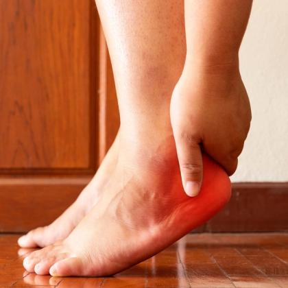
Plantar fasciitis is a common cause of heel pain, accounting for up to 15% of foot complaints requiring professional care and affecting both athletic and sedentary populations. It is best understood as a mechanically driven, degenerative condition resulting from repetitive loading that exceeds the plantar fascia’s capacity for repair, rather than a purely inflammatory process. Key risk factors include abnormal foot biomechanics, limited ankle dorsiflexion, posterior chain tightness, obesity and prolonged weight-bearing. In this episode, Dr Roger Henderson looks at the epidemiology and pathophysiology of plantar fasciitis, reviews key clinical features and examination findings, discusses differential diagnoses and appropriate investigations, and outlines evidence-based management strategies, with a focus on practical guidance for primary-care clinicians.
Key take-home points
- Plantar fasciitis accounts for up to 15% of all foot complaints requiring professional treatment and affects both athletic and non-athletic populations, with a lifetime prevalence of approximately 10%.
- The condition most commonly affects working adults aged 25–65 years, with higher prevalence in women, individuals with elevated BMI and those whose occupations involve prolonged standing or weight-bearing.
- Plantar fasciitis is best understood as a mechanically driven overuse condition in which repetitive loading exceeds the plantar fascia’s capacity for tissue repair.
- The plantar fascia plays a critical role in stabilising the longitudinal arch, and excessive tensile forces during gait contribute to microtrauma at its medial calcaneal origin.
- Biomechanical risk factors include pes planus, pes cavus, limited ankle dorsiflexion, excessive pronation or supination, prolonged standing, jumping activities and sudden increases in training intensity.
- Tightness of the gastrocnemius–soleus complex is a key and often under-recognised contributor, increasing plantar fascial tension during toe-off and impairing normal gait mechanics.
- Obesity, advancing age and heel fat pad atrophy further increase plantar loading by reducing the foot’s ability to absorb impact forces.
- Although plantar fasciitis can be associated with seronegative spondyloarthropathies, most cases occur without systemic disease; bilateral symptoms increase suspicion of an inflammatory cause.
- Calcaneal heel spurs are common incidental findings and do not cause plantar fasciitis, as many asymptomatic individuals have spurs and many symptomatic patients do not.
- Histological evidence demonstrates that plantar fasciitis is primarily a degenerative condition, characterised by collagen disorganisation and micro-tearing with minimal inflammation.
- Imaging findings on ultrasound and magnetic resonance imaging support a degenerative process, showing plantar fascia thickening, heterogeneity, calcifications and occasional partial tears.
- Classic symptoms include sharp inferomedial heel pain that is worst with the first steps in the morning or after rest, improves transiently with movement, and worsens with prolonged activity.
- The diagnosis of plantar fasciitis is clinical, and imaging or laboratory investigations are unnecessary unless symptoms are atypical, persistent or suggest alternative pathology.
- First-line management should focus on patient education, activity modification, stretching of the plantar fascia and calf muscles, appropriate footwear and other conservative measures.
- Most patients improve within 1 year, but recovery can be prolonged; recognising plantar fasciitis as a degenerative condition allows for realistic expectations and more effective long-term management.
Key references
- Rhim HC, et al. Life (Basel). 2021;11(12):1287. doi: 10.3390/life11121287.
- Koc Jr TA, et al. J Orthop Sports Phys Ther. 2023;53(12):CPG1-CPG39. doi: 10.2519/jospt.2023.0303.
- Dyck, DD, Boyajian-O’Neill, LA. Clin J Sport Med. 2004;14(5):305-9. doi: 10.1097/00042752-200409000-00010.
- Schwartz EN, Su J. Perm J. 2014;18(1):e105-107. doi: 10.7812/TPP/13-113.
- Cole C, et al. Am Fam Physician. 2005;72(11):2237-2242.
Create an account to add page annotations
Add information to this page that would be handy to have on hand during a consultation, such as a web address or phone number. This information will always be displayed when you visit this page